Smew Schmoo (and Other Histopathologic Works of Art)
As a pathologist at the San Diego Zoo, a large portion of my job is messy, ugly, bloody, and downright gross. Not that I mind that – it’s an occupational hazard when your job is observing and performing postmortem examinations. Death, though a natural part of life, is rarely pretty. However, microscopic examination is another story.
Tissues sampled from living animals to diagnose disease (i.e. biopsies) and from deceased animals as part of a necropsy (or postmortem examination) are processed for histopathology (for details, see this blog post). Histopathology involves microscopic examination of tissues to study manifestations of disease and is a standard diagnostic tool used by pathologists in human and veterinary medicine.
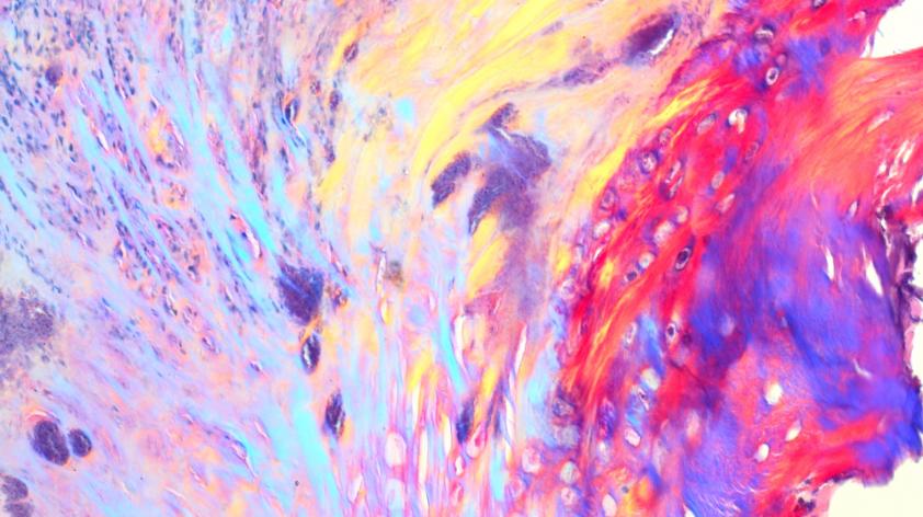
In contrast to “gross” pathology, the term aptly applied to macroscopic examination which only a pathologist is excited to look at, histopathology can often be considered visually appealing or even beautiful. Take for example a recent biopsy I examined from a smew (a type of duck). Clinically, this bird had an injury to its elbow that resulted in formation of a large scab or crust on its wing. Because this scab was impeding healing, clinicians removed it and submitted it for histopathologic examination. Diagnostically, it was not particularly interesting, consisting of the expected inflammatory response to damaged and infected tissue, but from an artistic perspective, it was spectacular!
As you can see from the images above and below, a stunning swirl of a rainbow of colors was revealed in the stained tissue under polarized light. Such a colorful image was definitely a highlight in a day of standard pinks and purples through the microscope. A couple other examples of “histo art” are included below. What can I say? Sometimes science and art are one and the same!


