


Parasite
June 01, 2020
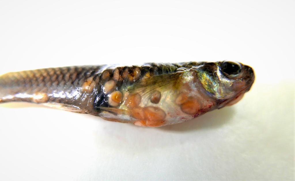
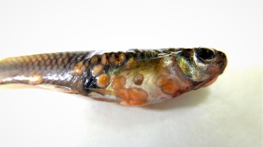
No, we aren’t talking about this year’s Oscar-winning film masterpiece, but the real thing. Sorry in advance to all you sushi lovers (including me)! The photo above and below shows a recent mosquitofish (Gambusia affinis) submitted to the Disease Investigations pathology service for diagnosis of some strange nodules all over its body.
These fish are not technically part of the San Diego Zoo collection but live in some of our freshwater aquatic habitats as biological vector controls. Mosquitofish are so named because they feed on mosquito larvae and thus help reduce populations of these annoying (and disease vectoring) pests. In fact, they are given out for free by the county of San Diego for this purpose!
This particular mosquitofish was one of many noted to have relatively large skin lesions within a pool providing habitat for other fish, reptiles, and native birds. As one of the most severely affected, this fish was caught and humanely euthanized for pathologic evaluation. As you can see in the image, there were a couple dozen tan to orange masses in this fish. Incision of a couple nodules allowed examination of the contents, which turned out to be – you guessed it – parasites.
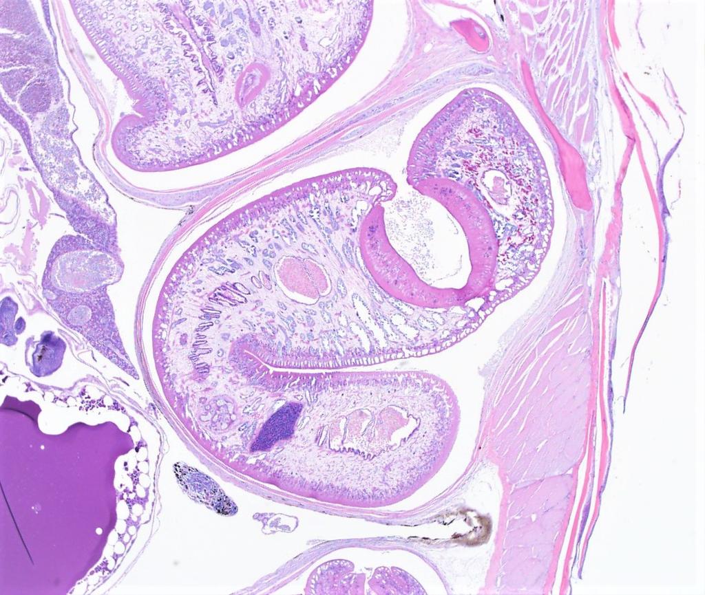
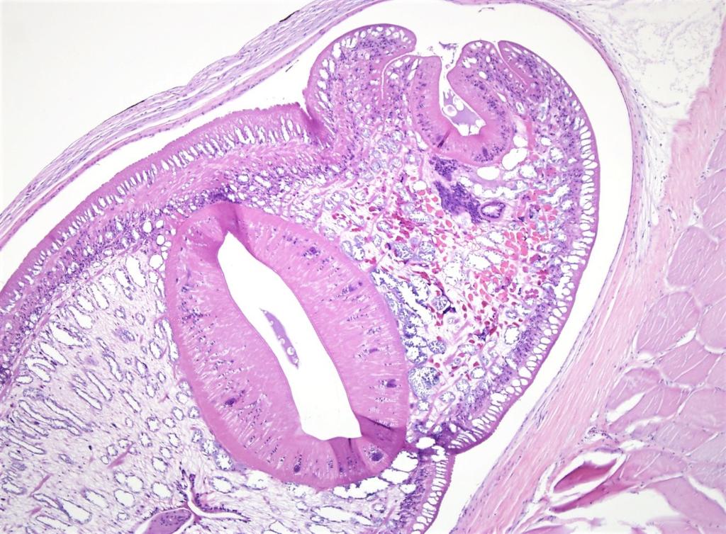
These particular parasites were larval stages of trematodes, also called flukes or flatworms, identifiable by their flattened, non-segmented, tapered body and oral and ventral suckers.
After examination of the fresh fish, we fixed it whole in formalin and processed it for histopathology. The additional images below show the parasites on histopathologic (microscopic) examination. In addition to being in the skin, the encapsulated trematode larvae were deeper in the body wall and within the lining of the body cavity.
This type of larval trematodiasis is not uncommon in fish due to the life cycle of these parasites. Although the exact type of trematode was not determined in this case, it would likely be one that in its adult form infects birds, mammals, reptiles, or other fish that prey on mosquitofish. Ingestion of the infected fish by the definitive host would allow the adult trematode to mature in the host’s intestinal tract and start producing eggs. The eggs are shed into the environment where they hatch and infect an invertebrate. After a time in the aquatic invertebrate, the next stage of the larval parasite leaves that host to infect a mosquitofish, which is then eaten by the final host to repeat the life cycle. It all seems needlessly complex, but such is the life of a parasite!
To interrupt this life cycle, veterinary clinicians decided to treat these fish with an anthelminthic drug that would kill the trematode larvae within the fish. Hopefully soon the native herons and egrets will be able to enjoy their sushi again, parasite-free!