


Putting eggs in a basket, step 1: Recover the eggs – Ovum Pickup (OPU) procedure
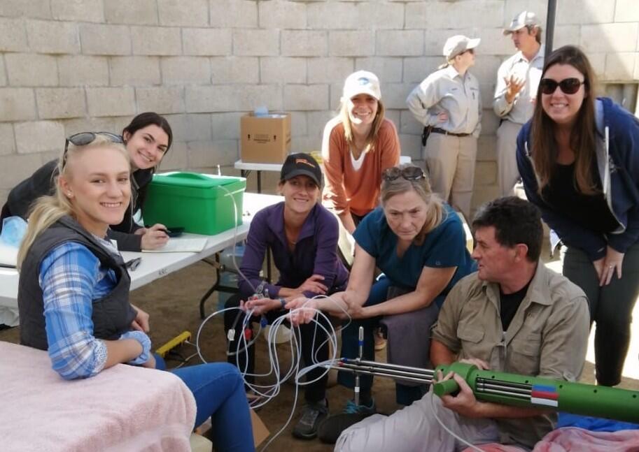
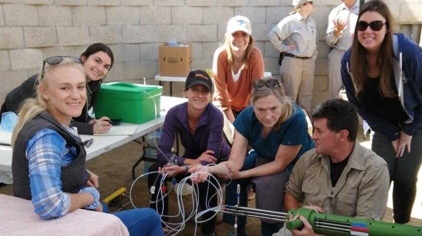
You may recall that I was fortunate enough to travel to South Africa to train on a procedure called ovum pickup (OPU) with our collaborator, Dr. Morné de la Rey. We recently performed this procedure on some of the females at the Rhino Rescue Center, with Dr. de la Rey so we could try our in vitro culture system and hopefully produce rhino embryos.
There is a lot of planning that goes in a procedure like this. First and foremost is the safety of the animal. This procedure does require anesthesia, which our veterinary services monitors very closely. But I want to talk a little about the technical aspects of the OPU procedure and how we get oocytes (ovum) in order to produce embryos in vitro.
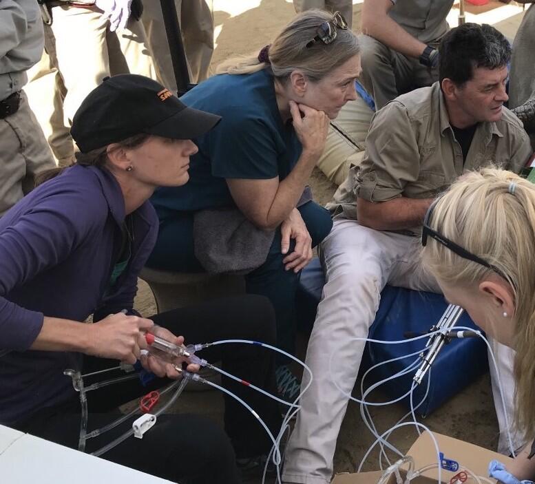
Once the rhino is comfortably asleep, a probe is positioned to begin collecting the oocytes. The probe is a specially designed piece of equipment that holds an ultrasound transducer (microconvex, to be specific) and the needles that will extend to ‘pickup’ the oocytes. The visual field that the ultrasound produces is aligned with the path that the needle will take, therefore this procedure is ‘ultrasound guided.’ We know that the needle will travel through the visual field of the ultrasound, which is why maintaining a visual of the ovary and follicle we intend to aspirate is so important. You may recall from previous posts that the follicle is the structure on the ovary that houses the oocyte. So, theoretically, there is an oocyte in every visible follicle on the ultrasound.
Next, we spend time taking care to recover the entire contents of the follicle, and hopefully the ooctye within it. To do this we use a double lumen needle (basically, a needle within a needle). The outer needle is connected to a source of flush medium while the inner needle is connected to a collection vessel that is also attached to an aspiration unit that creates negative pressure. The outer needle pushes fluid into the follicle, the inner needle sucks fluid out. When the needle is inside a follicle, the aspiration pump is turned on to suck out the fluid; simultaneously, fluid is pushed into the follicle create turbulence. This turbulence hopefully dislodges the oocyte and allows it to be sucked out with the rest of the fluid. The coolest part is that all of this can be seen on the ultrasound screen. Once the follicle has been sufficiently flushed and aspirated for a final time, not leaving any fluid in the follicle, the needle is retracted and aligned with a different follicle, and the process repeated.
This process is completed on as many follicles as can be safely accessed on both ovaries. All of the fluid that has been sucked out is now in the collection vessel – hopefully it has the oocytes as well! There are several considerations to make during this complex procedure: first, the animal’s safety, but also the viability of the eggs. We warm the medium that is flushed into the follicle as well as the collection vessel that holds the fluid removed from the follicle. The medium we use to flush the follicle is formulated to nourish the oocytes the collection vessel is glass so the eggs do not stick to the walls, and the pressure we aspirate the fluid with is carefully adjusted so the oocytes are not damaged as they travel to the collection vessel.
Once the procedure is finished, the collection vessel is immediately taken to the lab where the fluid is searched for the eggs so that they can be cultured and hopefully become an embryo after fertilization. That is a fascinating process as well, and I will leave that for Carly Young to tell you about. I will say that the procedure was a success! We recovered oocytes from all four animals that we aspirated – even a whopping 13 from RRC females Livia!
We are so excited to see this procedure safely recover oocytes from these females and look forward to what it will enable us to do.
Video below: This video shows the ultrasound image of the ovary during an OPU procedure, in this case equine. The large dark circles are follicles, each contains an egg. Because the ultrasound transducer and needle are aligned, when follicles are positioned in the 'field of view' of the ultrasound, the needle has a predictable path. In this video, the follicle is positioned in the upper left corner of the ultrasound screen. The needle will then enter the top left corner and puncture the follicle, the follicle collapses immediately, its contents are aspirated - and hopefully the egg!
Note: the video orientation is correct, the ultrasound unit is on its side to aid visual follicle and needle alignment.