


A Seminal Case
July 30, 2019
Working as a veterinary pathologist for San Diego Zoo Global means that on a regular basis I get up close and personal with many species I’ve never seen before (though unfortunately usually after they are deceased). So, being confronted with an unfamiliar species is not unusual, but it is rare for an anatomic structure to catch me off guard, as recently happened with a fairly common native species.
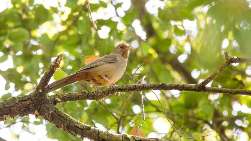
At the end of May, I was presented with a wild male California towhee. This bird was found dead on zoo grounds and was submitted for necropsy (animal autopsy) as part of our disease surveillance program. We perform postmortem exams on all wildlife that die at our facilities in order to screen for diseases that could affect other native wildlife, our collection animals, or the public.
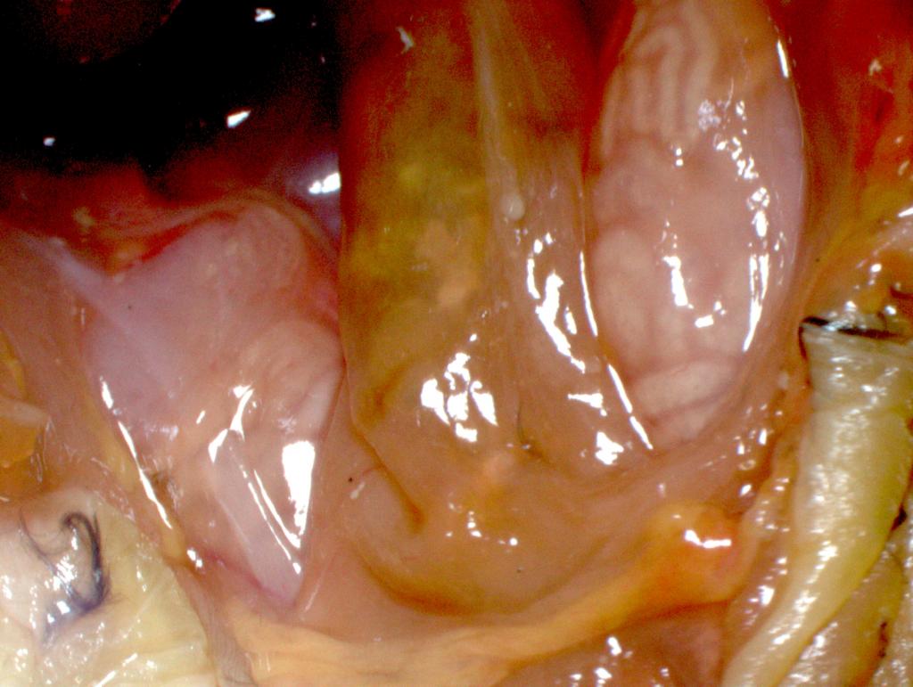
This particular bird was in suboptimal nutritional condition and had a prominent swelling in the region of the vent (the opening of the cloaca) with crusting of the surrounding skin. This swelling corresponded internally to two tan, approximately 10 mm long by 5 mm diameter structures on either side of the cloaca (Figure 1).
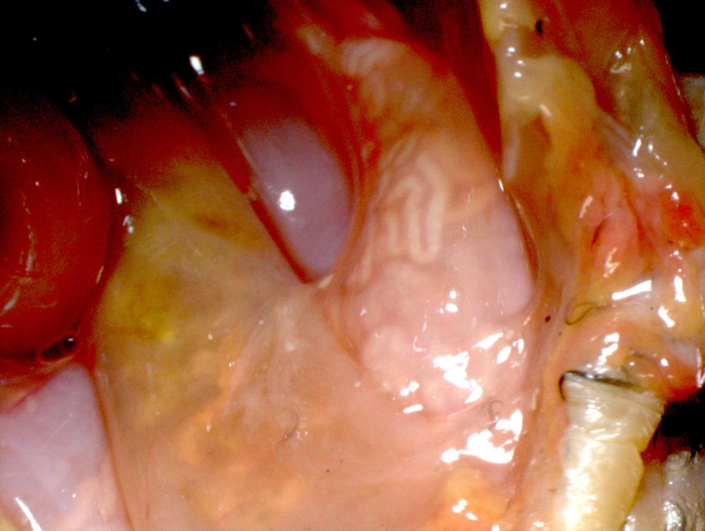
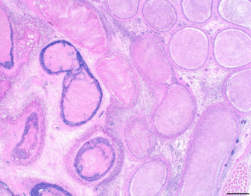
These structures appeared to have an internal network of whitish, serpentine tubular structures up to 1 mm in diameter visible through a thin wall (Figure 2). In 10 years of performing necropsies on birds, I could not remember ever previously noticing structures like these!
So, were they normal or abnormal? As it turns out, they were a little bit of both!
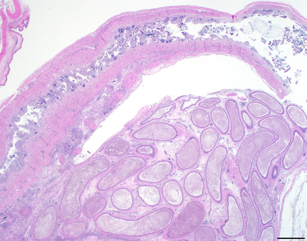
After consulting various avian anatomy texts, especially those providing detail about the male reproductive tract, which opens into the cloaca along with the digestive tract and urinary tract, I was able to determine that each of these structures is a seminal glomus. Seminal glomera are convoluted tubular expansions of the ductus deferens, the structure that transports sperm from the testes to the cloaca (Figure 3).
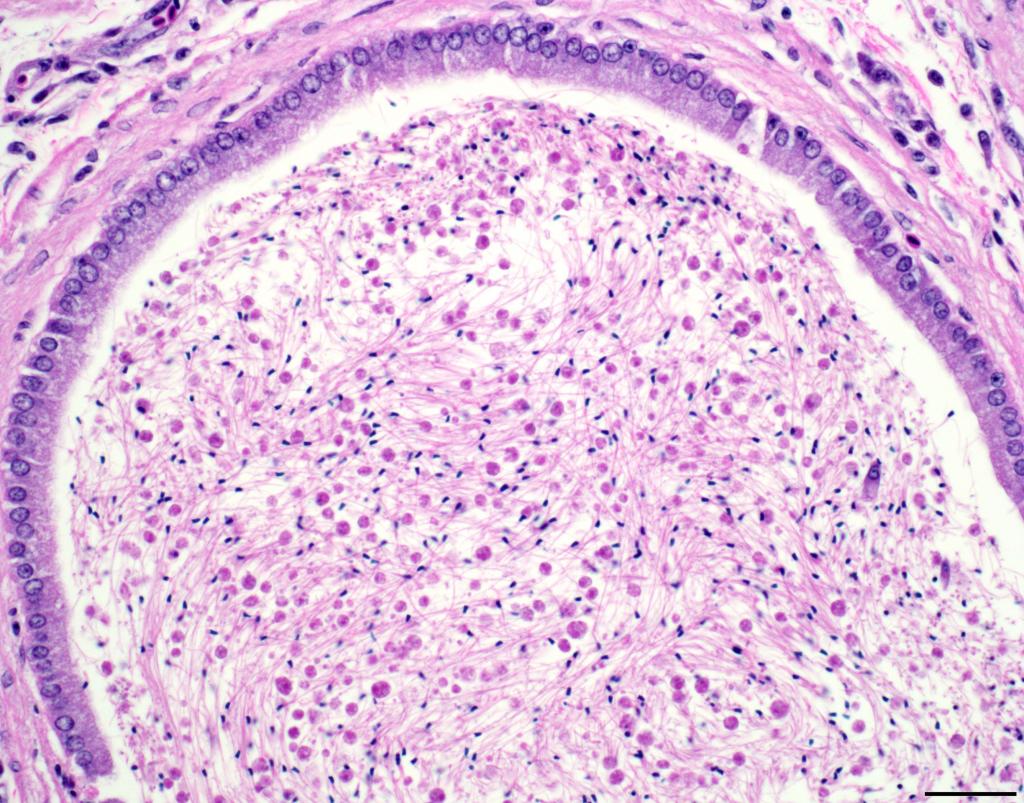
Each seminal glomus serves as a storage site for sperm (Figure 4), and therefore they enlarge during the breeding season when the bird is reproductively active. They are most prominent in passerines (songbirds), and the resulting bulging of the cloaca (called a cloacal promontory) and protrusion of the vent can be used by field biologists to determine that a bird is a male.
Although the anatomic structure is normal, the seminal glomera and vent region of this particular towhee were not.
The crusting around the vent was an area of ulceration and bacterial infection that involved the adjacent cloaca and seminal glomera (Figure 5). The infection and inflammation could have obstructed the ducts and caused even greater enlargement. It is likely that the bacterial infection was the cause of death in this bird.
As a pathologist, sometimes I may not pay enough attention to a tissue until it contains a pathologic condition or lesion, and I’m sure this won’t be my last anatomy lesson! Now if only I knew what the medical term for “inflammation of the seminal glomus” was – glomeritis, anyone?