Ultrasonography: A window into rhino reproduction
In the ongoing fight against extinction, the Reproductive Sciences team does our part with many different species. For Reproductive Sciences, one of our main focuses is on assisted reproductive techniques for the southern white rhino (SWR) in the hopes that we can bring its closest relative, the northern white rhino back from the brink of extinction.
One of the most important tools we have is the ability to perform routine, un-sedated ultrasound exams on the resident SWRs Helene, Wallis, Amani, and Victoria at the Nikita Khan Rhino Rescue Center (RRC) to complement the hormone data we gather from fecal samples. In a collaborative effort, ultrasound exams are performed by Dr. Parker Pennington and Dr. Barbara Durrant while hormone assays are performed by Marissa Taylor.
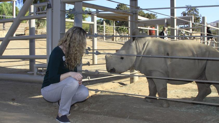
At the RRC, located at the San Diego Zoo Safari Park, we have some of the best trained SWRs in the world. Our wildlife care specialists have worked tirelessly to form relationships with the females who call the RRC home to ensure they can soar through medical exams, blood draws, and weekly ultrasounds. In fact, the RRC wildlife care specialists can even ask the SWRs to urinate and defecate in preparation for procedures and rectal ultrasound exams.
Thanks to the time spent forming relationships and bonds with the RRC rhinos, ultrasound exams run very smoothly. The rhino comes into a yard, urinates and defecates, and waits patiently to be let into the ultrasound area. Once she is in place, she starts chowing down on her favorite fruits, veggies, pellets, and hay while the reproductive science team gets to work.
First, there is a quick check to ensure all fecal matter is out of the way. If there is more, it is gently removed as it can obstruct the view generated by the ultrasound. Next, a small ultrasound probe is inserted into the rectum and the connected ultrasound screen shows us images of her reproductive tract.
Rhino ultrasound exams to visualize ovaries cannot be performed vaginally or abdominally as in other species due to the location of the reproductive tract within the female. Luckily, the angle of the ultrasound probe within the rectum of the rhino allows for a bird's eye view of the majority of the reproductive tract.
Once we have an image, we get to work locating her uterine horns and her ovaries. We take photographs and videos of the same locations on the reproductive tract each time in order to compare any changes from one exam to the next. The process takes only a few minutes, but the information we obtain is extremely valuable.
When the ultrasound exam is complete, we look at the photographs and videos and take measurements of uterine horn size, follicle (a spherical structure within the ovary that contains an oocyte) number, follicle size, and level of uterine edema (the collection of fluid within the tissue). We take note of how these factors changed from the last exam, how long ago that exam was, and try to predict what will happen next (e.g. ovulation).
We also track these changes over time and from cycle to cycle. This information allows us to compare what we see real time in the ultrasound images with the cycle pattern information we obtain through retrospective fecal progesterone monitoring. Further, we can generate patterns for an individual and predict what she is likely to do.
For example, Wallis is likely to ovulate a large follicle on her own, whereas Helene is not – and therefore we use hormone treatments to induce Helene to ovulate.
In the case of Amani and Victoria, based on real time and historical information, ultrasound images confirmed the right time to induce ovulation and when to artificially inseminate. This led to Edward and Future, our first calves born from artificial insemination in North America!
Without the countless hours of bonding and training by the wildlife care specialists, the reproductive sciences team would not have been able to generate ultrasound information for the RRC rhinos – and would never have led to welcoming Edward and Future! We are so proud to be a part of this interdisciplinary team and can’t wait to see what milestones are next.

