


In vitro – What does it mean?
We have blogged a lot about the work we do in the lab at the San Diego Zoo Safari Park. The Reproductive Sciences team is constantly seeking ways to ensure the survival of species. We concentrate on certain focal species, but ideally, we would have the techniques and technology to preserve them all.
One way we are working toward this goal is by developing in vitro culture techniques. You may have already read about this several times from Carly Young, Nicole Ravida, Elena Ruggeri, Barbara Durrant, and myself. I want to take a moment to explain what we call ‘in vitro culture’ as a process in general.
While 'in vitro' technically means 'in glass', it is widely used to mean 'outside the body'. Many people first heard of in vitro work in reference to the human fertility field, where the term ‘test tube baby’ was coined. The general concept is to produce an embryo in the lab that would be capable of becoming a fetus and eventually a newborn. There are limitations to these techniques, and several critical steps along the way.
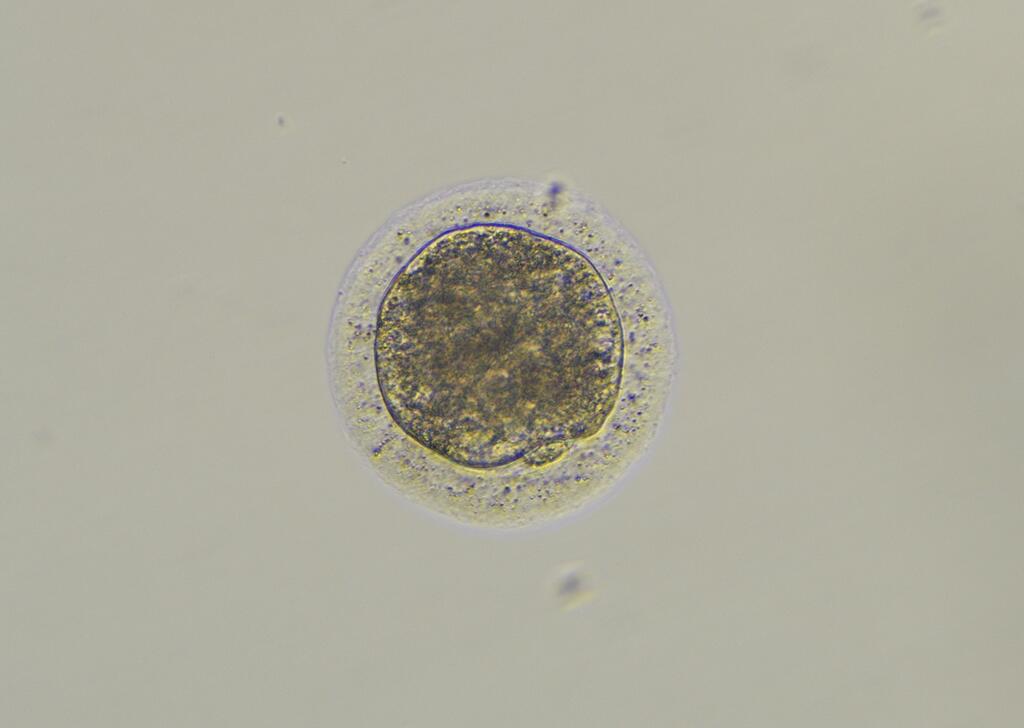
But, very simply put, it starts with a sperm and an egg and ends with an embryo called a blastocyst. The blastocyst is the last stage of embryo development that we can support in the lab (in vitro). After that, the blastocyst must either be frozen for long term storage or transferred to a surrogate for gestation. If all goes well, she will carry the pregnancy to term and deliver a healthy offspring. To culture animal embryos we try to mimic what the body would be providing during embryo development. We formulate a medium with similar components and an environment (temperature and humidity) that is similar to the conditions inside the body. The ‘medium’ is the liquid mix of hormones, sugars, growth factors and protein that the oocytes and embryos will develop in. Each stage is carefully formulated to provide the necessary nutrients at the correct stage. Throughout the process we also need a well-controlled environment, which is where incubators come in. We adjust the temperature, humidity and air gasses inside the incubators to replicate the conditions inside the body.
The time it takes to go from sperm and egg to blastocyst is quite short, between 5 and 11 days, depending on the species. It is important to remember that these are general rules, and that every species has their own specific requirements to achieve critical milestones in the first days of development.
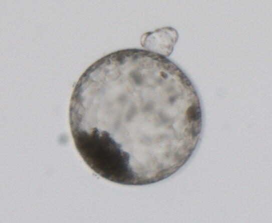
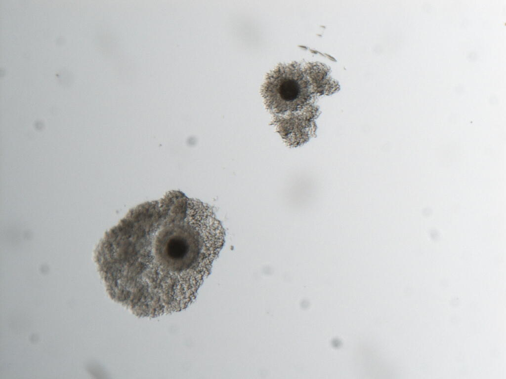
The first milestone is the for the egg to become ‘mature.’ This means that the egg is capable of being fertilized. It is evidenced by the extrusion (pushing out) of a polar body. This signals that the egg is metabolically active and can now accommodate a sperm…should one arrive. In the body (in vivo), maturation would occur just before ovulation (in most species).
Once the egg has matured, it has a time limit - a life span - so sperm must find it in time. The specific components and factors needed to support maturation in vitro vary by species, but this is a critical step. Without it, the egg cannot develop further and will not create an embryo. Since this process would typically take place in a follicle on the ovary, we try to provide it with similar hormone concentrations and conditions….if we know them!
If the egg matures, it is then introduced to sperm for fertilization. This may be by simply placing a number of sperm in a drop of medium in a petri dish with the egg. We refer to this as in vitro fertilization or IVF. It is the simplest way to produce fertilized eggs – let the sperm do the work! In some species, this process does not work, and we don’t always understand why. But, we can get around it by using micromanipulators (exactly what they sound like) to hold a single egg and inject a single sperm, a process called intracytoplasmic sperm injection (ICSI; pronounced ICK-see). This method of fertilization is used routinely in human fertility clinics.
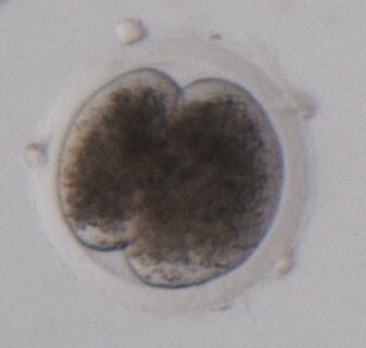
Once the sperm and egg have been combined (one way or another), next comes the difficult task of waiting. Waiting to see if the fertilization was a success, waiting to see if the sperm and egg are now one entity – an early embryo. The first indication is called ‘cleavage’. One cell cleaves into two. If those cells continue to divide (as they must to create a whole creature), they become 4 cells, 8 cells, 16, 32, and so on. The embryo begins to change shape on the inside, creating a space in the middle, with all the cells pushed to the perimeter. At this point we have a blastocyst! Please check out this time lapse video of blastocyst development.
The blastocyst is the final step that can be supported in vitro. Each of these steps takes place over mere days and requires a different medium, a different environment. Remember, we are trying to mimic the body. So, in vivo maturation takes place in the ovary, which has its own medium. In vivo fertilization takes place in fallopian tubes, with its own medium. Embryo development normally takes place in the uterus, with its own medium. We recreate each of these in a dish, with media and incubators for the in vitro process, gently moving the precious orbs from drop to drop as they develop. The whole concept…and process is an incredible thing.
All Photos courtesy of Carly Young.